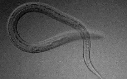
An overview of the mechanisms by which parasitic worms successfully invade and survive within their hosts.
In an article published as a part of the plos pathogens pearls - a collection of short educational articles on the latest topics concerning pathogen research - authors Boyett and Hseih describe the five most versatile interactions between helminthic parasites and their host tissues. The following is an excerpt from the article 'Wormholes in Host Defense: How Helminths Manipulate Host Tissues to Survive and Reproduce', reproduced here under the creative commons attribution license.
"A major tool in the helminth arsenal is the ability to compromise the host epithelium, which is the first line of defense against parasitic infection. For instance, schistosomes encounter numerous epithelial barriers in their life cycle. These barriers include the skin during its invasion by cercariae, the lung during schistosomular transit, and the intestine and bladder during egress of schistosome eggs in the fecal and urinary streams, respectively. Based on in vitro studies, schistosomal cercariae are thought to employ proteases such as cercarial elastase to degrade human skin elastin and collagen during the first stage of infection, allowing penetration of skin epithelium. When schistosome worms mature, mate, and lay eggs, it becomes essential for the eggs to exit the host body, as the parasite life cycle will not continue without a secondary host. Schistosoma haematobium eggs, in particular, have developed molecular strategies for easing passage from the bladder tissue to the urinary stream. Mouse bladders injected with S. haematobium ova exhibit decreased transcription of all uroplakin genes and various tight junction related genes. The structural proteins that these genes encode are essential for the maintenance of bladder epithelial integrity and the sequestration of urine from tissues extraneous to the bladder. The altered transcription of these genes may benefit helminth survival by facilitating access to the bladder lumen and the next stage of the life cycle.
In contrast to schistosomes, or blood flukes, which aggressively invade their host through the skin, many gastrointestinal helminths traverse host epithelia after being ingested. The whipworm, Trichuris trichiura, may alter host intestinal epithelial cells using secretory proteins. It is postulated that these proteins enable the worm to form ion conduction pores in the lipid bilayer of cecal epithelial cells, allowing the parasite to burrow its head into a nutrient-rich, intracellular microenvironment, and providing shelter from the mechanical movements of gut tissue."
Read further at http://tinyurl.com/padkqgk
Cover image: Necator Americanus (Wikipedia).
Advertisement
Stats
- Recommendations n/a n/a positive of 0 vote(s)
- Views 1112
- Comments 0